
Columbia University
Irving Medical Center
Neurological Institute
710 West 168th Street, 3rd floor
(212) 305-1818
Featured Research
12th ANNUAL TAUB INSTITUTE
RESEARCH RETREAT
November 15, 2022
Zebrafish as a Functional Genomics Tool for Alzheimer's Disease
Caghan Kizil, PHD
Associate Professor of Neurological Sciences (in Neurology and the Taub Institute)
The molecular mechanisms of actions of the genes that are clinically relevant to the pathogenesis of Alzheimer’s disease (AD) in humans are not fully known. Therefore, reductionist vertebrate animal models to investigate the biological relevance of AD-related genes are necessary. We are using a genetically tractable model organism, zebrafish. With its ease of genetic manipulation, high similarity of gene/protein structures to humans, advanced live imaging techniques and feasibility of high-throughput genetic or behavioral screens, zebrafish is a useful model organism,
We generated an adult zebrafish model of amyloid toxicity by cerebroventricular injection of human amyloid-beta42 monomers into the cerebrospinal fluid of the fish brain and with stable expression of human genes. Using histological analyses, imaging, transcriptomics and phenotypic screens, we determined the similarities of zebrafish amyloid pathology to humans. Zebrafish induce microglial response, synaptic degeneration, neuronal death, and impairment in cognitive abilities that resemble human brains phenotypically and molecularly. We identified several molecular mechanisms that underlie the pathology-induced neuro-immune response in zebrafish and translated this finding to mammalian models. From a large clinical functional genomics approach through a genome-wide association in AD patients in relation to cerebrovascular risk factors, we identified that the gene FMNL2 links cerebrovascular disease and AD, by regulating the astroglial and blood vessel interactions and controlling the efficient clearance of toxic proteins from the brain. We are following this route by investigating clinically identified candidate genes for their biological activity in vivo by performing CRISPR/Cas9-mediated gene editing and single cell transcriptomics. Additionally, we are using zebrafish as a pre-clinical drug screening tool by synthesis of original multitarget ligands and developing design-based novel hit compound identification for synaptic integrity.
Function and Role for Plasma Membrane-Bound Neuroproteasomes
Kapil Ramachandran, PHD
Assistant Professor of Neurological Sciences (in Neurology, Neuroscience, and the Taub
Institute)
For decades, we have defined proteasomal degradation as the action of a series of ubiquitylating enzymes and the 26S proteasome, a large complex which recognizes ubiquitylated substrates and degrades them. Recently, we have described a new variant of the proteasome in neurons, which is bound to the neuronal plasma membrane, lacks the 19S cap, and degrades substrates co-translationally. Moreover, we find that neuroproteasomes degrade proteins across the membrane into released extracellular peptides, which act as a new principle of cell-cell signaling. Here, will we describe our progress on understanding the function of these novel complexes in the brain.
CNS Immune Surveillance in AD and Other Neurodegenerative Diseases
Wassim Elyaman, PHD
Assistant Professor of Neurological Sciences (in Neurology, the Taub Institute, and the
Institute for Genomic Medicine)
Alzheimer’s disease (AD) is an age-related neurodegenerative disease leading to dementia. The central nervous system (CNS) is continuously monitored by immune cells, many of which circulate through the cerebrospinal fluid (CSF) and the meninges. The CNS must balance between vigilantly detecting for potentially harmful factors and resolving any immunological responses that in themselves can create damage if left unabated. The meninges surrounding the CNS host diverse populations of immune cells that influence how CNS-related immune responses develop. Recent studies demonstrate that meningeal lymphatics play a role as a communicator between the brain and peripheral immunity. The etiology of AD is currently unknown, but a number of diverse risk factors have been identified, that increase susceptibility to AD. Some of these risk factors are infectious agents. In fact, the “pathogen hypothesis” suggests that AD pathogenesis may be triggered and/or exacerbated by an infectious agent. Viral, fungal, and bacterial pathogens have all been associated with AD, including herpes simplex virus type 1 (HSV-1), human herpesvirus 6A (HHV-6A), HHV-7, cytomegalovirus (CMV), as well as the intracellular bacteria Chlamydophila pneumoniae and Porphyromonas gingivali. Our hypothesis reasons that analyzing meningeal T-cell repertoire in AD would provide novel data about microbial-specific T-cells. Here, we provide evidence of microbial antigens in AD meninges using fresh autopsies provided by the New York Brain Bank (NYBB) at Columbia University Medical Center. Through single-cell RNA coupled with T-cell receptor (TCR)-sequencing of immune cells in the AD meninges, we identified microbial-specific TCR clonotypes, many of which were hyperexpanded with 25-50% of the TCR repertoire. Analysis of cell-cell communications revealed signaling pathways that are involved in promoting clonal T cells. Our study supports the association between infectious agents and AD pathogenesis. Ongoing studies are aimed to analyze the effects of microbial-specific T-cells on CNS inflammation.
Crossing the Blood Brain Barrier: the Holy Grail of Drug Delivery for Neurodegenerative Disease
Carol Troy, MD, PHD
Professor of Pathology and Cell Biology and Neurology (in the Taub Institute)
Despite great effort by many researchers over many years, we do not yet have effective therapies for Alzheimer’s Disease and other neurodegenerative diseases. While there is very promising research identifying potential therapeutic targets, we also need to consider the methods of delivery that maximize the efficacy of nascent therapies and minimize potential adverse effects. The primary obstacle facing therapeutic development is crossing the blood-brain barrier. There are many delivery modalities available for delivering therapies, from oral to intraparenchymal, each with its own advantages and drawbacks. Minimally invasive routes such as oral delivery are comfortable for patients, but their penetrance into the brain is uncertain. On the other hand, intraparenchymal delivery is guaranteed to enter the brain, but is incredibly invasive and introduces risk of infection. We have previously utilized intranasal instillation of cell permeant peptides which provided morphologic, cellular and functional protection in rodent models of acute cerebral ischemia. We are now determining whether combining intranasal instillation with focused ultrasound will provide maximal efficacy of cell permeant therapeutics in chronic neurodegeneration.
New Approaches to Integrate Methylation, Histone Modification, and Gene Expression
QTLs for Alzheimer's Disease Gene Mapping
Gao Wang, PHD
Assistant Professor of Neurological Sciences (in Neurology and the Gertrude H.
Sergievsky Center)
Statistical fine-mapping seeks to determine genetic variants responsible for complex traits, given evidence of an association of a genomic region with highly correlated variants due to linkage disequilibrium. In the recent years, fine-mapping has been widely applied to GWAS to identify putative causal variants in complex traits including Alzheimer's disease (AD). Although molecular quantitative trait loci (QTL) such as eQTLs have been successfully integrated to fine-map GWAS loci, integration of QTLs for spatially distributed molecular phenotypes such as DNA methylation (mQTL) and histone modification (haQTL) remains a challenge. In particular, these phenotypes contain numerous correlated measurements within a genomic region (eg CpG sites, histone modification peaks), making it difficult to pinpoint the exact QTLs regulating one or multiple such phenotypes while accounting for LD between these QTLs. Currently, no attempts have been made to systematically fine-map mQTLs and haQTLs. To integrate these molecular phenotypes to AD GWAS, we developed a novel approach to fine-map spatially correlated high-dimensional phenotypes. We implemented a variable selection model we have previously developed into a non-parametric regression models using wavelets. We developed a flexible mixture prior combined with an Empirical Bayes approach to fit a wide variety of spatial and temporal data. Our new method, fSuSiE, can efficiently fine-map thousands of CpG sites simultaneously, and the output is easy to interpret. As an application, we have fine-mapped mQTL and haQTL from the Religious order Study and the Memory and Aging Project (ROSMAP). We are currently extending our work to integrate with other QTLs as well as AD GWAS loci for joint statistical fine-mapping.
Unraveling Mechanisms of Stress-Induced Tau Pathology
Clarissa Waites, PHD
Associate Professor of Pathology and Cell Biology and Neuroscience
Prolonged exposure to high levels of glucocorticoids (GCs), the main stress hormones, damages the brain and is a risk factor for Alzheimer’s disease (AD). Here, we investigate the mechanistic underpinnings of two major drivers of GC-induced neurotoxicity, mitochondrial dysfunction and Tau pathology, in murine hippocampus. We find that GCs induce mitochondrial damage by promoting the opening of the mitochondrial permeability transition pore (mPTP) via upregulation of its activating subunit, cyclophilin D. Interestingly, inhibition of mPTP opening in vitro protects against GC-induced mitochondrial dysfunction as well as Tau phosphorylation and oligomerization, suggesting that Tau pathology occurs downstream of mitochondrial damage. Moreover, inhibiting mPTP opening in vivo is protective against Tau pathology, synaptic and neuronal loss, and behavioral deficits induced by GCs. These findings show that mPTP opening is a precipitating factor in GC-induced mitochondrial dysfunction and Tau pathogenesis, and suggest that mitochondria are promising therapeutic targets for mitigating stress-related brain pathology.
Friend and Foe: Stress Signaling in Neurodegeneration
Ulrich Hengst, PHD
Associate Professor of Pathology and Cell Biology (in the Taub Institute)
The integrated stress response (ISR) is activated in many neurodegenerative conditions including Alzheimer’s disease. The primary goal of cellular stress signaling is to reestablish homeostasis for example through the adaption of protein synthesis and energy expenditure. On the other hand, if the stress response is insufficient or too pronounced it might lead to cell death. This dual role of the cellular stress response, being potentially both pro-survival and pro-degenerative, has confused efforts to target it therapeutically, with both inhibitors and activators of the ISR being proposed and investigated. Here, we suggest concentrating instead on effector pathways of the ISR that are either primarily pro-survival or pro-degenerative. As an example for a pathology-driving stress response pathway we present the AD-linked heterodimerization of two stress response transcription factors, ATF4 and CREB3L2. The formation of this heterodimer is sufficient to explain around 50% of gene expression changes in AD brain and to induce AD-typical cellular changes, including the dysregulation of the retromer, altered APP processing, and tau hyperphosphorylation. We discuss strategies to prevent or correct the effects of ATF4-CREB3L2 heterodimerization. As an example for a resilience-boosting stress response pathway we report our discovery of a novel cell non-autonomous stress signal, TAILS, which is generated in response to various stressors, including in the AD brain, and greatly increases the cellular survival in the face of those stressors. We find that TAILS enhances SHH signaling, thereby increasing mitochondrial mass and respiration and cellular redox potential. The therapeutic potential of TAILS is established by our finding that its administration completely prevents cell death and preserves hippocampal function in a 4-vesicle occlusion model of global ischemia. Together, our data support our hypothesis that targeting ISR effector pathways rather than the ISR itself is a promising strategy in neurodegeneration.
Using Spatial Genomics to Study the Central Nervous System in Health and Disease
Hemali Phatnani, PHD
Assistant Professor of Neurological Sciences (in Neurology)
My research program focuses on using novel tools and technologies in conjunction with cellular and animal models and patient-derived tissue samples to understand how disease-causing mutations perturb the intricate interplay between glial and neuronal cells in ALS-FTD. To understand the role of intercellular interactions in disease, we apply spatially-resolved transcriptomic and proteomic approaches to deconvolve both spatial and cell-type specific changes in gene expression across entire brain or spinal cord regions from rodent and human post-mortem tissue. Using such an integrated, cell ensemble-resolution, multi-omic approach, our studies generate multidimensional datasets that will enable us to determine cell- and region-specific molecular correlates of functional impairment in ALS-FTD. Moreover, they may provide a platform for other investigators to unveil markers specific to their disease of study. This will be encouraged and facilitated by sharing our platform and data with the broad scientific community.
KEYNOTE LECTURE
Complex Roles of Proteins with Low Complexity Intrinsically Disordered Domains in
Normal Neurobiology and Neurodegeneration
Peter St George-Hyslop, OC, FRS, FRSC, FRCPC
Professor of Neurology (in the Taub Institute)
Until recently, little was known about the biological role of domains found in many proteins that have low amino acid complexity and no intrinsic fixed structure (termed “low complexity domains” or “intrinsically disordered domains”). However, in the last several years, it is become clear that proteins containing these domains have the unique thermodynamic property of being able to reversibly phase transition from mono-dispersed states in solution into more condensed states (e.g. liquid: liquid droplets, hydrogels, glasses etc). Crucially, these intrinsically disordered proteins can co-partition with other similarly structurally disordered molecules (e.g. other proteins, nucleic acid polymers) to form transient “membraneless organelles”. These membraneless organelles include nucleoli, P bodies, stress granules, neuronal transport granules, and play key roles in diverse cell biological processes including transcription, translation and signal transduction. It has also become apparent that mutations in these proteins can alter their biophysical properties, promoting accelerated condensation into stable, fibrillar aggregates associated with disease.
The early analyses of proteins with such intrinsically disoreded domains led to the now well-accepted understanding of how reversible condensation of RNA binding proteins (e.g. FUS) underpins their role in axonal transport of selected mRNAs to distal neuronal compartments. However, these early studies also led to questions about how these transient, flexible, liquid droplets, which lacked attachments to motor proteins, could be transported down the crowded axoplasm without breaking-up under the shear stress. We show that, for some RNP granules, this is accomplished by using ANXA11 as a molecular tether to allow hitchhiking of the liquid droplet RNP granule on the surface of lysosomes. This work also uncovered the unexpected the existence of crosstalk between membraneless organelles and classical membrane-bound organelles. Based on these observations, we suggest that there may be rich bidirectional interactions (termed “phase coupling”) between the cell biology of membraneless and membrane-bound organelles. These interactions may drive both the clustering of cell membrane lipid rafts and receptors (driven by sub-membrane cytoplasmic biomolecular condensates) and the transient assembly of sub-membrane cytoplasmic signalling complexes (driven by the membrane receptor proteins).
The induction of such sub-membrane condensates by cell surface receptor activation is likely to be a feature of signal transduction in many CNS cell types, not just neurons where much of the work on intrinsically disordered proteins has so far focused upon. An unpublished example will be discussed.
Contact Us
630 West 168th Street
P&S Box 16
New York, NY 10032
Phone: 212 305-1818
Fax: 212 342-2849
Email: taubinstitute@columbia.edu
Brain Donation
Our ability to understand Alzheimer's disease is dependent upon studying brain tissue.
For more information, please contact Donovan Laing at (212) 305-9086 dal2190@cumc.columbia.edu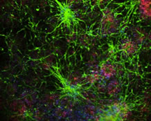
This website uses cookies as well as similar tools and technologies to understand visitors' experiences. By continuing to use this website, you consent to Columbia University's usage of cookies and similar technologies, in accordance with the Columbia University Website Cookie Notice.